Skip to document

Cc 0 orientation - EKG and Hemodynamics

EKG and Hemodynamics
Course

Critical Care (408)

18 Documents
Students shared 18 documents in this course
Academic year: 2020/2021
Uploaded by:
0followers
14Uploads
25upvotes

Comments

Please sign in or register to post comments.
  • MG
    Awesome notes, thank you!

Related Studylists

MEDICALcolorful guides211

Preview text

Normal Breath Sounds inspiration > expiration low pitch soft no adventitious sounds

Vesicular Breath Sound most of lung field low pitch soft & short exhalation long inhalation

Bronchovesicular Breath heard over main bronchus upper right posterior lung field medium pitch exhalation = inhalation

Bronchial Breath Sound trachea only high pitch loud and long exhalation

Crackles excessive secretions/fluid in airway short, discrete popping/crackling sounds associated w/ PNA, pulmonary edema, pulmonary fibrosis, atelectasis

Wheezes vibrations when air flows at fast speed through narrow airway high-pitched, squeaking, whistling sound associated w/ asthma or bronchospasm

Pleural Friction Rub produced by irritated pleural surfaces rubbing together creaking, leathery, dry, coarse, sound associated w/ pleural effusion, pleurisy

Absent no airflow to particular portion of lung associated w/ pneumothorax, pneumonectomy, lung mass, complete airway obstruction

Diminished little airflow to particular portion of lung sound intensity is reduced associated w/ atelectasis, COPD, obesity

Stridor sign of obstruction in trachea or larynx loud, high-pitched sound (sounds like snore) wake pt. up to r/o stridor  if awake & still has snoring sound  positive for stridor Chest Tube Nursing Actions Chest Tube always keep drainage system below level of chest & free of kinks troubleshoot cause if there’s change in previous assessment if cause can’t be confirmed  notify physician ALWAYS have sterile Vaseline gauze on hand in case of accidental dislodgement if chest tube comes off  apply pressure on insertion site w/ sterile Vaseline gauze and call physician

Value of ScvO remove fluid & air from pleural space three chest tube settings suction water seal (to gravity) clamped assessed if pt. can tolerate function without chest tube or if they don’t need chest tube anymore air leaks bubbling in the drainage system assess and identify cause every shift

central venous oxygen saturation normal: 70 – 80% ScvO2 can be used as convenient surrogate to a PA catheter for detection & rapid TX of tissue hypoxia sustained ScvO2 <70% associated w/ higher mortality

Arterial Line (A-line, ABP) Central Venous Catheter (triple lumen CVC, CVL)

Pulmonary Artery Catheter

continuous monitoring of ABP non-traumatic sampling of arterial blood gas (ABG) do NOT infuse

continuous monitoring of central venous pressure monitoring of central venous oxygen saturation (ScvO2)

reserved for most hemodynamically unstable patients for diagnosis & evaluation of heart dz, pulmonary HTN, shock

medications/IV in A-line Mean Arterial Pressure (MAP) normal: 70-100mmHg mean > 60 to perfuse vital organs MAP = [ 2(diastolic) + systolic] ÷ 3 arterial waveform in EKG dicrotic notch (aortic valve closing  little back pressure bump) & appropriate waveform for each QRS highest point = SBP, lowest point = DBP

fluid/volume status monitoring, blood draws, meds, IV fluid Central Venous Pressure measures right atrial pressure (PRELOAD)  reflects force-filling of R atrium & R ventricle measured at junction of vena cava and Rt atrium & CVP only measured w/ central line unable to transduce CVP from peripheral or short PICC line normal: 2-5 - indicate fluid balance

states

normal values PAP: 20-30mmHg/5- 10mmHg measured w/ balloon deflated PAOP: 5-12mmHg wedge pressure w/ balloon inflated left ventricle at end diastolic pressure (LVEDP) CO: 4-6L/min

Zeroing a Line level transducer @ phlebostatic axis (4th ICS, MAL) close stopcock to patient open transducer to air press “zero” on monitor/touchscreen close transducer to air open stopcock to patient quick flush of line (square test)

Nursing Care on A- Line, CVC, PA maintain pressure bag at 300mmHg maintain gravity infusion at 3mL/hr change heparin/saline flush solution bag q24h or when it runs out change pressure tubing q48-72h (2-3days) per unit protocol assess site & change dressing per protocol assess if pressure readings & waveforms are normal or abnormal run EKG strips of waveform & place on chart every shift

Central Line Complications phlebitis at insertion site air embolism hematoma arrhythmia catheter malposition late complications thrombus formation infection

External Ventricular Device (EVD) measures ICP & drain CSF indicated in intracranial hemorrhage, traumatic brain injury, bacterial meningitis most common/important lifesaving procedures in neuro ICU subarachnoid screw EVD normal: 4-15mmHg slightly elevated: 15- 20mmHg if >20mmHg for 10mins  NOTIFY PROVIDER

Heart Sounds S1 (lubb) mitral & tricuspid valves (AV) close high pitched “lubb” sound loudest at apex beginning of systole isometric contraction

S

aortic & pulmonic valves (semilunar) close high pitched “dubb” sound loudest at base end of systole & beginning of diastole isometric

S

rapid filling of ventricles in early diastole low pitched Kentucky | “what the heck” normal in children and pts w/ high ↑ CO pregnant mom, athletes) pathological in

S

sign of diastolic heart failure (late diastole) Tennessee | bad joke drum active ischemia chronic HTN ( syllables – S4) left ventricular hypertrophy (LVH)  impaired relaxation

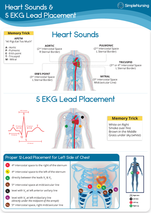
aortic = 2nd ICS on Rt chest pulmonic = 2nd ICS on Lt chest erb’s point = 3rd ICS tricuspid = 4th/5th ICS mitral = 5th ICS all people enjoy touching me

decrease to 0/min for 18hrs

Was this document helpful?

Cc 0 orientation - EKG and Hemodynamics

Course: Critical Care (408)

18 Documents
Students shared 18 documents in this course
Was this document helpful?
Normal Breath
Sounds
inspiration >
expiration
low pitch
soft
no adventitious
sounds
Vesicular Breath
Sound
most of lung field
low pitch
soft & short
exhalation
long inhalation
Bronchovesicular
Breath
heard over main
bronchus upper
right posterior lung
field
medium pitch
exhalation =
inhalation
Bronchial Breath
Sound
trachea only
high pitch
loud and long
exhalation
Crackles
excessive secretions/fluid
in airway
short, discrete
popping/crackling sounds
associated w/ PNA,
pulmonary edema,
pulmonary fibrosis,
atelectasis
Wheezes
vibrations when air flows at
fast speed through narrow
airway
high-pitched, squeaking,
whistling sound
associated w/ asthma or
bronchospasm
Pleural Friction Rub
produced by irritated pleural
surfaces rubbing together
creaking, leathery, dry,
coarse, sound
associated w/ pleural
effusion, pleurisy
Absent
no airflow to particular
portion of lung
associated w/ pneumothorax,
pneumonectomy, lung mass,
complete airway obstruction
Diminished
little airflow to particular
portion of lung
sound intensity is reduced
associated w/ atelectasis,
COPD, obesity
Stridor
sign of obstruction in
trachea or larynx
loud, high-pitched sound
(sounds like snore)
wake pt. up to r/o stridor if
awake & still has snoring
sound positive for stridor
Chest Tube Nursing Actions Chest
Tube
always keep drainage
system below level of chest
& free of kinks
troubleshoot cause if there’s
change in previous
assessment
if cause can’t be confirmed
notify physician
ALWAYS have sterile
Vaseline gauze on hand in
case of accidental
dislodgement
if chest tube comes off
apply pressure on insertion
site w/ sterile Vaseline
gauze and call physician
Value of ScvO2
remove fluid & air from
pleural space
three chest tube settings
suction
water seal (to gravity)
clamped
assessed if pt. can
tolerate function
without chest tube or if
they don’t need chest
tube anymore
air leaks
bubbling in the drainage
system
assess and identify cause
every shift
central venous oxygen
saturation
normal: 70 – 80%
ScvO2 can be used as
convenient surrogate to a PA
catheter for
detection & rapid TX of
tissue hypoxia
sustained ScvO2 <70%
associated w/ higher
mortality
Arterial Line (A-line, ABP) Central Venous Catheter
(triple lumen CVC, CVL)
Pulmonary Artery Catheter
continuous monitoring of
ABP
non-traumatic sampling of
arterial blood gas (ABG)
do NOT infuse
continuous monitoring of
central venous pressure
monitoring of central
venous oxygen saturation
(ScvO2)
reserved for most
hemodynamically unstable
patients for diagnosis &
evaluation of heart dz,
pulmonary HTN, shock